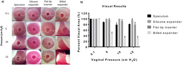
Fig 6.
a) Results from experimental determination of percent visual area showing images of the mock cervix (with centered os) in the vaginal phantom with the graves speculum, silicone expander, flat tip inserter and the billed expander. Images are captured at different vaginal pressures 0.1, 5, 10 and 15 cm H2O. b) Grouped bar plot of mean percent visual area of the cervix for the different devices under the different pressures. Error bars are standard deviations.

