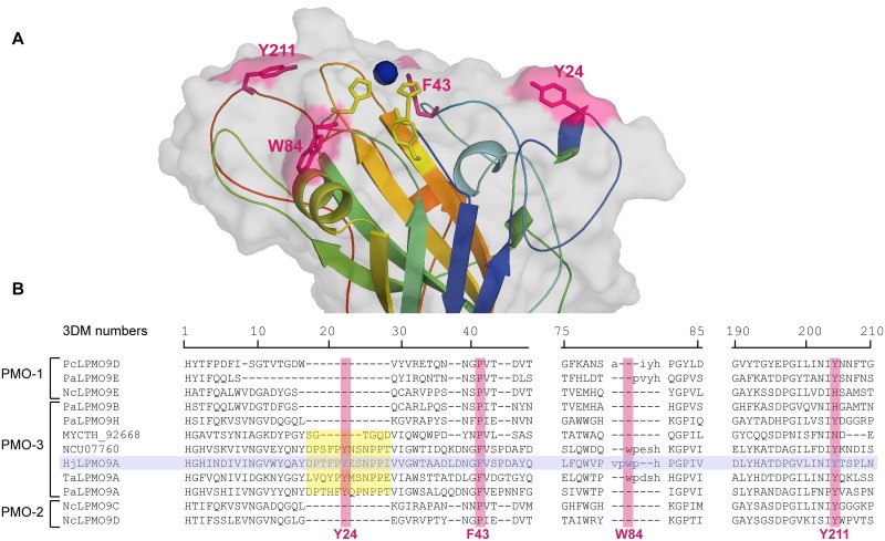
Fig 5. Aromatic surface residues in the C1/C4-oxidizing HjLPMO9A.
(A) Homology model of HjLPMO9A (based on 3ZUD as template) with the aromatic surface residues selected for alanine scanning in pink stick representation. Active site residues are shown as yellow sticks, the copper ion as a blue sphere. (B) 3DM structure based multiple sequence alignment [40] of AA9 characterized LPMOs with known regioselectivity. The residues aligned with the Y24, F43, W84 and Y211 aromatic surface residue of HjLPMO9A are highlighted in pink. Residues in the 3DM core alignment are represented by capitals, the alignment of structurally variable regions are in lower case. The insertion typical for most C1/C4-oxidizing LPMOs is marked in yellow.

