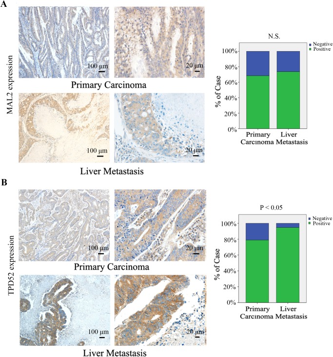
Fig 2. Immunohistochemical staining for MAL2 and TPD52 in human CRC tissues.
(A,B) Expression levels of MAL2 and TPD52 in primary carcinoma tissuesand liver metastasis tissues from patients with CRC were detected by immunohistochemical staining (×100, 100 μm; ×400, 20 μm). MAL2 and TPD52 expression levels were quantified as shown as the right bar charts. *P<0.05 compared with primary carcinoma; N.S., no significance.

