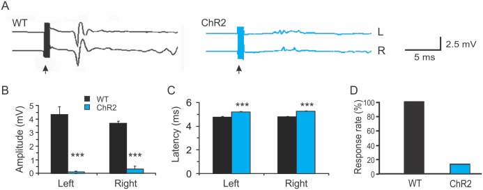
Fig 1. Reduced motor evoked potentials induced by transcranial magnetic stimulation in Thy1-ChR2 transgenic mice.
A. Representative traces showing bilateral TMS-evoked motor evoked potentials (MEPs) in a wild type C57 Black mouse (WT; black) and a Thy1-ChR2 transgenic mouse (blue). The black arrows indicate times of transcranial magnetic stimulation (TMS). B-D. The amplitudes of bilateral TMS-evoked MEPs were dramatically decreased in the ChR2 mice compared with WT mice (B); the latencies of bilateral TMS-evoked MEPs were significantly increased in the ChR2 mice (C), and the percentage of mice in which MEPs were induced by TMS was greatly reduced in the ChR2 mice (D) (***: p<0.001, Student t-test).

