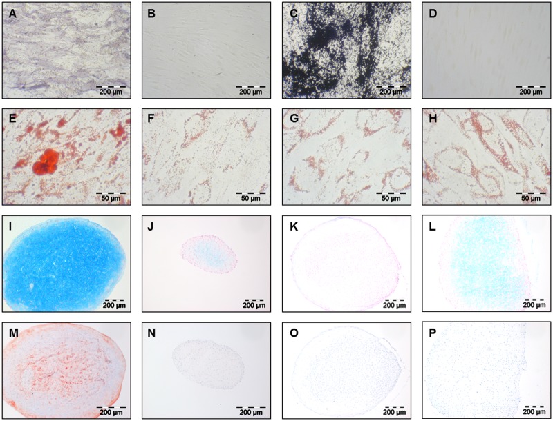
Fig 2. Histological and immunochemical stainings of osteo-, adipo- and chondrogenically induced clonal cultures.
Alkaline phospahtase staining of osteogenically induced clonal cultures (A) and uninduced contols (B); Von Kossa staining of osteogenically induced clonal cultures (C) and uninduced contols (D); Oil red O staining of adipogenically inducible (E) and non-inducible (G) clonal cultures and corresponding uninduced controls (F,H); Alcian blue staining of chondrogenically inducible (I) and non-inducible (K) clonal cultures and corresponding uninduced controls (J,L); Collagen Type II immunochemical staining of chondrogenically inducible (M) and non-inducible (O) clonal cultures and corresponding uninduced controls (N,P); A-D and I-P 100x magnification, E-H 400x magnification.

