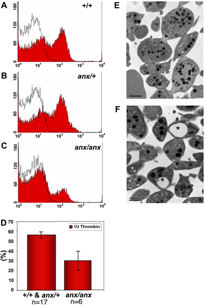
Fig. 2.

anx/anx mice show deficits in secondary platelet aggregation. (A-C) The effect of (A) +/+, (B) anx/+ and (C) anx/anx platelets on CD62 (P-selectin) expression, a marker for α-granule secretion, was detected using flow cytometry. After activation with thrombin, CD62 levels on the platelet surface were significantly reduced in (C) anx/anx platelets. Black lines represent resting platelets and red shading denotes thrombin-stimulated platelets. Representative examples of at least four independent experiments are shown. (D) The percentage of secondary platelet aggregation in +/+ and anx/+ animals was 56.4±3.2% (n=17) and anx/anx animals 23.9±9.8% (n=6) (Mann–Whitney Test, P=0.026). Data represented as mean±s.e.m. (E,F) Electron microscopy analyses revealed that (F) anx/anx platelets appeared normal, but often contain large vacuoles (indicated by asterisks) absent in (E) +/+ platelets. The abnormal vacuoles were observed in 17.5±5.9% of anx/anx platelets (counted 57-63 platelets per replicate (n=4).
