Fig. 3.
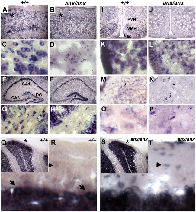
Tyro3 RNA localization in +/+ and anx/anx brains at P21. RNA in situ hybridization detected Tyro3 RNA (A) in layers 2/3, 5 and 6 of +/+cortices. (B) Cortices of anx/anx mice show diffuse mutant (C19T) Tyro3 RNA expression. (C) At high magnification, in the +/+ cortex, Tyro3 is localized in soma at the base of processes. (D) In anx/anx mice, C19T-Tyro3 RNA localization appears disorganized in the soma of cortical neurons. In the hippocampus of (E) +/+ and (F) anx/anx mice, Tyro3 RNA is present in CA1, CA3, and the dentate gyrus (DG). (G) At higher magnification, Tyro3 is expressed in cells within the granule cell and polymorphic layers of the +/+ DG. (H) In anx/anx, only a few cells in the granule cell and polymorphic layers of the DG express C19T-Tyro3. (I) In the +/+ hypothalamus, Tyro3 is expressed in the median eminence, arcuate nucleus (ac), and ventromedial hypothalamus (VMH), and at lower levels in the paraventricular nucleus of hypothalamus (PVN). (J) In the anx/anx hypothalamus, C19T-Tyro3 expression appeared so diffuse at low magnification that it is difficult to distinguish it in specific subregions. At higher magnification, (K) in the +/+ arcuate nucleus, Tyro3 RNA is localized in processes emerging from neuronal soma, but (L) in the anx/anx arcuate nucleus, C19T-Tyro3 RNA localization appears disorganized. Tyro3 is expressed in neurons within the dorsal raphe nuclei of (M) +/+ and (N) anx/anx mice. At high magnification, Tyro3 RNA is seen at the edges of (O) +/+ neuronal soma and in their emerging processes, but appears disorganized in (P) anx/anx neurons within the raphe nuclei. (Q) In the +/+ cerebellum, Tyro3 is expressed in granule cells and Purkinje cells, shown at higher magnification in (R) (marked with arrows). In the cerebellar molecular layer, Tyro3-expressing processes can be seen at high magnification. Tyro3 RNA faintly outlines the soma of cells – likely cerebellar stellate and basket cells (marked with arrowheads in R). (S) In anx/anx cerebella, C19T variant Tyro3 RNA is present in granule and Purkinje cells. (T) At higher magnification, mutant Tyro3-positive cell bodies (marked with arrowheads), which are likely stellate or basket cells, can be seen in the molecular layer. Mutant Tyro3-expressing processes are not apparent. Asterisks in A,B,E,F,I,J,M, N,Q and S indicate regions shown at higher magnification directly below each respective panel in C,D,G,H,K,L,O,P,R and T.

