
Fig. 3.
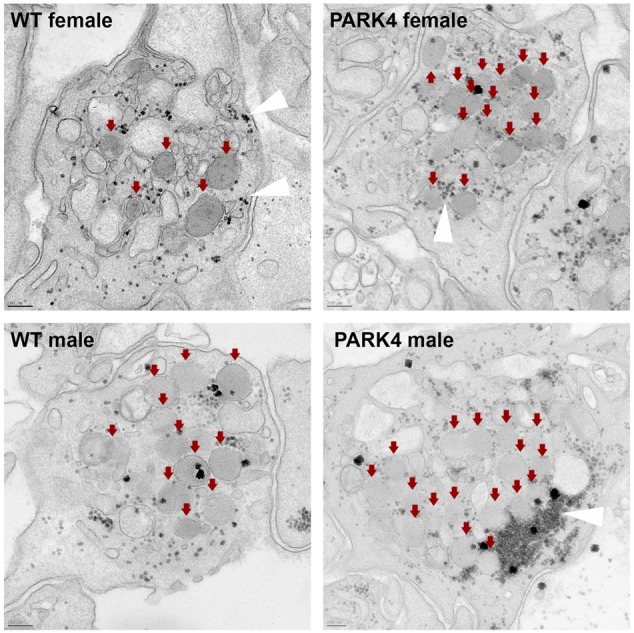
Electron microscopy of blood platelets after stimulus-triggered degranulation is depicted, illustrating centrally clustered α-granules (red arrows) and glycogen granules (white arrowheads), but no detectable protein aggregates in PARK4 cases versus matched WT relatives. Scale bars: 200 nm (n=2 control versus n=2 PARK4 individuals). WT, wild type.