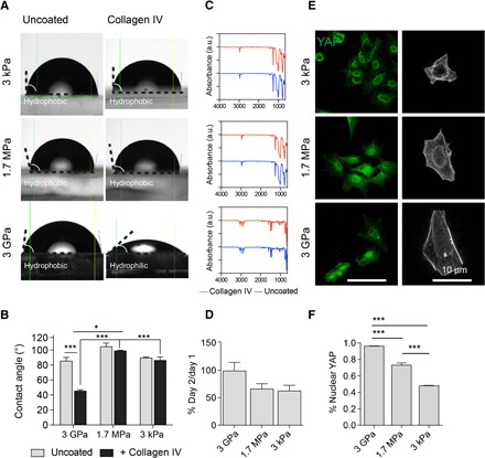
Fig. 1. PDMS substrates are permissive to hiPSC differentiation.

(A) Representative images of water droplets on collagen IV–coated and uncoated PDMS and E ~ 3 GPa substrates for contact angle measurements. (B) Contact angle quantification across substrates. (C) FTIR analysis of substrates with and without collagen IV coating. a.u., arbitrary units. (D) Attachment efficiency across substrates. (E) Sparse seeding elicits varied YAP (green) localization and spreading [filamentous actin (F-actin) in gray] dependent on substrates with corresponding (F) quantification. All data are presented as means ± SEM. *P < 0.05; **P < 0.01; ***P < 0.001. At least three replicates were performed.
