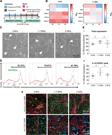
Fig. 4. Stiffness-primed mesoderm induction in serum-free conditions results in robust EC differentiation.

(A) Schematic of stiffness-primed mesoderm induction followed by EC differentiation on polystyrene plates. (B) Left: Gene expression of mesodermal markers for cells differentiated on soft 3-kPa substrates, normalized to expression from E ~ 3 GPa surfaces. Right: Gene expression analysis of cells differentiated on stiff 1.7-MPa substrates, normalized to expression from E ~ 3GPa surfaces. Color key is presented in log10 scale. (C) Bright-field images of cobblestone endothelial colonies (white arrows) on day 12 EVCs. (D) Representative day 12 EVC flow cytometry plots of VECad expression in red, with corresponding HUVEC VECad expression in green. (E) (i) Total VECad expression as a function of substrate stiffness. (ii) Percentage of VECad expression relative to HUVEC control samples. (F) Representative immunofluorescence images of day 12 EVC expression (top: green, eNOS; red, F-actin; blue, nuclei, bottom: green, vWF; red, CD31; blue, nuclei). Data are represented as means ± SEM. *P < 0.05, **P < 0.01, and ***P < 0.001, paired Student’s t test. At least three biological replicates were performed.
