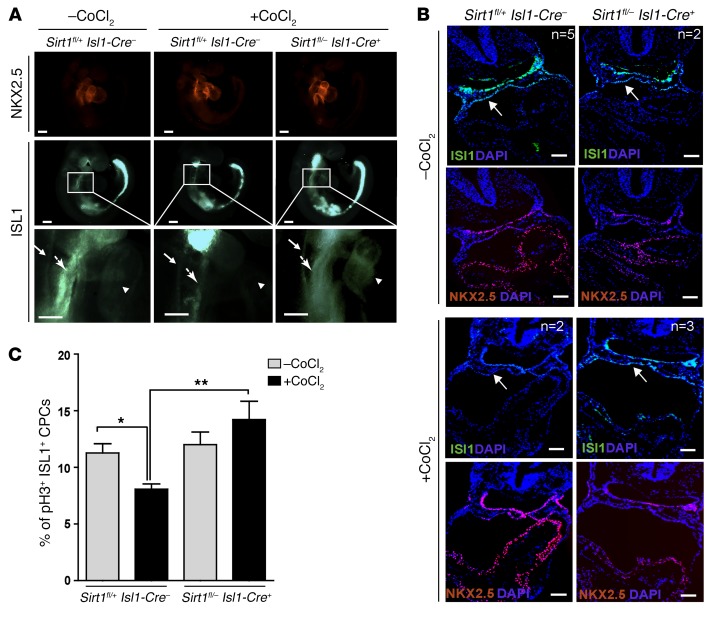
Figure 5. Inactivation of Sirt1 in ISL1+ cells enhances Isl1 expression and promotes proliferation of ISL1+ cells after induction of hypoxia responses.
(A) Whole-mount immunostaining of E9.5 control and Sirt1 mutant embryos for NKX2.5 and ISL1 with and without chemical induction of hypoxia responses (30 mg/kg body weight CoCl2 treatment). The pale green staining in the head and tail is due to unspecific autofluorescence. Neural tube (arrows with small arrowheads), cardiac mesoderm (arrows with big arrowheads), and heart tubes (arrowheads) are indicated. Embryos with or without chemical induction of hypoxia responses were obtained from 3 different litters. Scale bars: 100 μm. (B) Representative images of sections from control and Sirt1 mutant E9.5 embryos following whole-mount immunostaining for ISL1 and NKX2.5 with and without chemical induction of hypoxia responses (15 mg/kg body weight CoCl2 treatment). Embryos with (Sirt1fl/+ Isl1-Cre–: n = 2 Sirt1fl/– Isl1-Cre+: n = 3) or without (Sirt1fl/+ Isl1-Cre–: n = 5; Sirt1fl/– Isl1-Cre+: n = 2) chemical induction of hypoxia responses were obtained from 2 different litters. Note reduced ISL1 levels in the cardiac mesoderm (white arrows) in control, but not in Sirt1fl/– Isl1-Cre+, littermates after chemical induction of hypoxia responses. Scale bars: 200 μm. (C) Quantification of ISL1+ cell proliferation by pH3 immunostaining in control (–CoCl2: n = 4; +CoCl2: n = 5) and Sirt1fl/– Isl1-Cre+ E9.0 embryos (n = 3) after chemical induction of hypoxia responses. Note that inactivation of Sirt1 in the SHF rescues reduced proliferation of ISL1+ cells. *P < 0.05; **P < 0.01, ANOVA with Dunnett’s post hoc correction.

