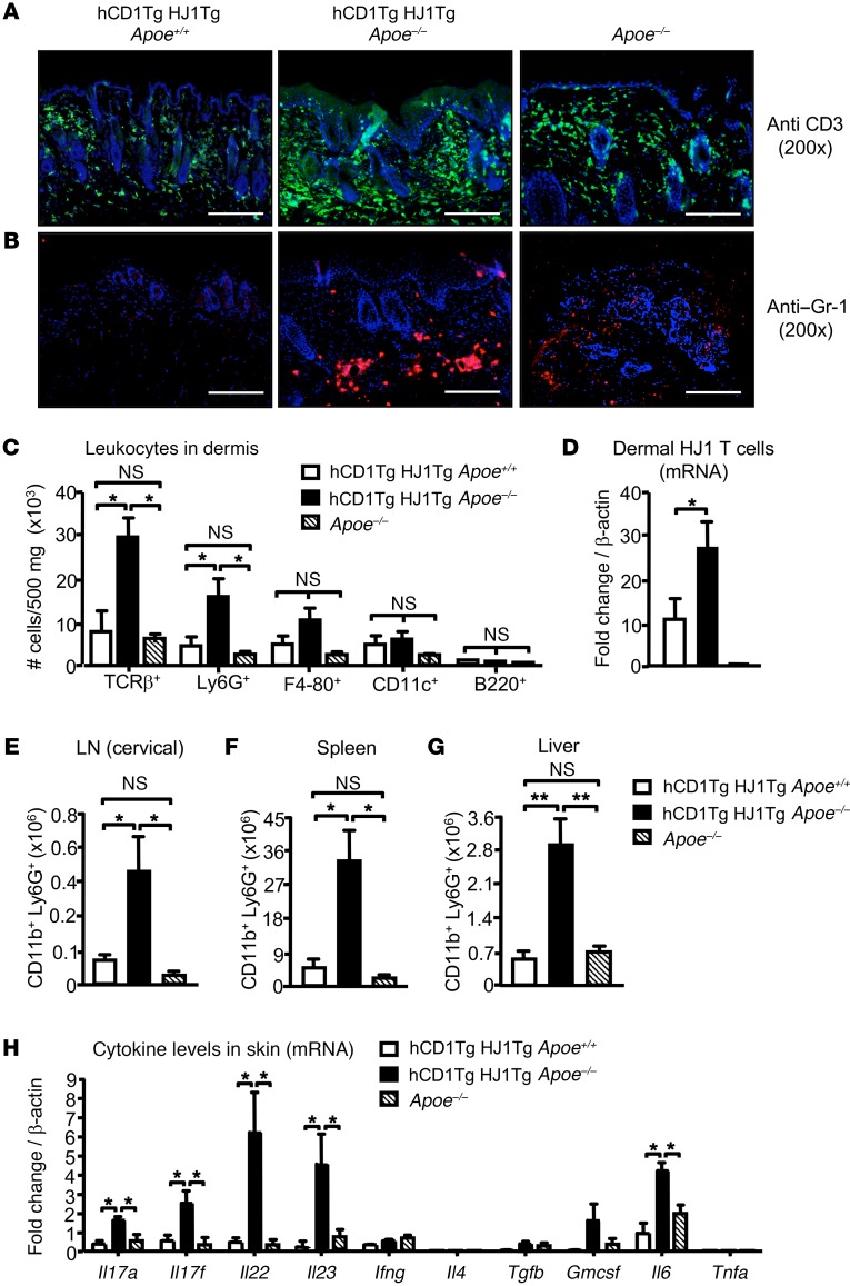
Figure 2. Diseased skin in hCD1Tg HJ1Tg Apoe–/– mice is characterized by T cell and neutrophil infiltrates.
Indicated organs were harvested from mice at about 25 weeks of age when hCD1Tg HJ1Tg Apoe–/– mice exhibited fulminant disease. (A and B) Immunofluorescence staining of skin sections from indicated mice with anti-CD3 (A) and anti–Gr-1 (B). Scale bars: 100 μm. (C) Bar graph depicts the absolute number of various leukocyte subsets in the dermis of mice, performed using flow cytometry (n = 3–5). (D) mRNA analysis of HJ1 T cells in the skin of the mice using HJ1 TCR–specific primers. (E–G) hCD1Tg HJ1Tg Apoe–/– mice have systemic neutrophil infiltration. Quantification of neutrophils (CD11b+Ly6G+cells) in the cervical LNs (E), spleen (F), and liver (G) was performed using flow cytometry (n = 4–6). (H) mRNA detected in the skin of different mouse strains using cytokine-specific primers (n = 3–5). mRNA levels were normalized relative to β-actin. Values are mean + SEM. ***P < 0.005; **P < 0.01. Statistical analyses were performed using 1-way ANOVA followed by Bonferroni’s post-hoc test.

