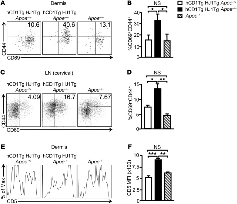
Figure 3. Increased activation of T cells is observed in the skin and cervical LNs of hCD1Tg HJ1Tg Apoe–/– mice.
Dermal and LN cells were isolated from indicated mice at 25 weeks of age, stained with anti-CD45 Abs, and gated on TCRβ+ cells. Cell-surface staining was performed for T cell activation markers. (A and C) Representative FACS plots of the expression of T cell activation markers CD69 and CD44 in the dermis (A) and cervical LNs (C). (B and D) Bar graphs depict the mean + SEM of the percentages of CD69+CD44+ T cells in the dermis (B) and LNs (D) of the mice (n = 5–10). (E) Representative FACS plots of CD5 expression on dermal T cells from indicated mice. (F) Bar graph depicts the MFI of CD5 expression on dermal T cells in different mouse strains (n = 3). ***P < 0.005; **P < 0.01; *P< 0.05. Statistical analyses were performed using 1-way ANOVA followed by Bonferroni’s post-hoc test.

