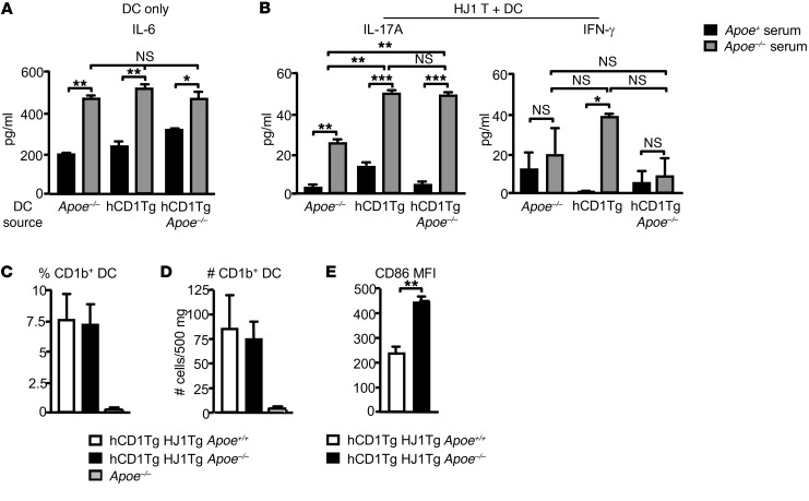
Figure 6. DC function and phenotype under hyperlipidemic conditions.
(A) Pam3Cys-treated hCD1Tg, hCD1Tg Apoe–/–, and Apoe–/– LN–derived DCs were cultured in Apoe+/+ and Apoe–/– mouse serum. IL-6 was measured by cytometric bead array (CBA) after 48 hours. (B) hCD1Tg, hCD1Tg Apoe–/–, and Apoe–/– DCs were cocultured with hepatic HJ1 T cells from hCD1Tg HJ1Tg Rag–/– mice in Apoe+/+ and Apoe–/– serum for 48 hours. ELISA was used to measure IFN-γ and IL-17A secretion. Data are representative of at least 2 independent experiments. (C–E) Cells from the dermis were isolated, stained with various Abs, and gated on CD45 and CD11b+CD11c+ DCs. Percentages (C) and numbers (D) of CD1b-positive cells in the skin were quantified. (E) CD1b+DCs (gated on CD11b+CD11c+ population) were examined for their expression of costimulatory molecule CD86 (n = 4). ***P < 0.005; **P < 0.01; *P < 0.05. Statistical analyses were performed using 1-way ANOVA followed by Bonferroni’s post-hoc test for 3 group comparisons and Student’s t test for 2 group comparisons.

