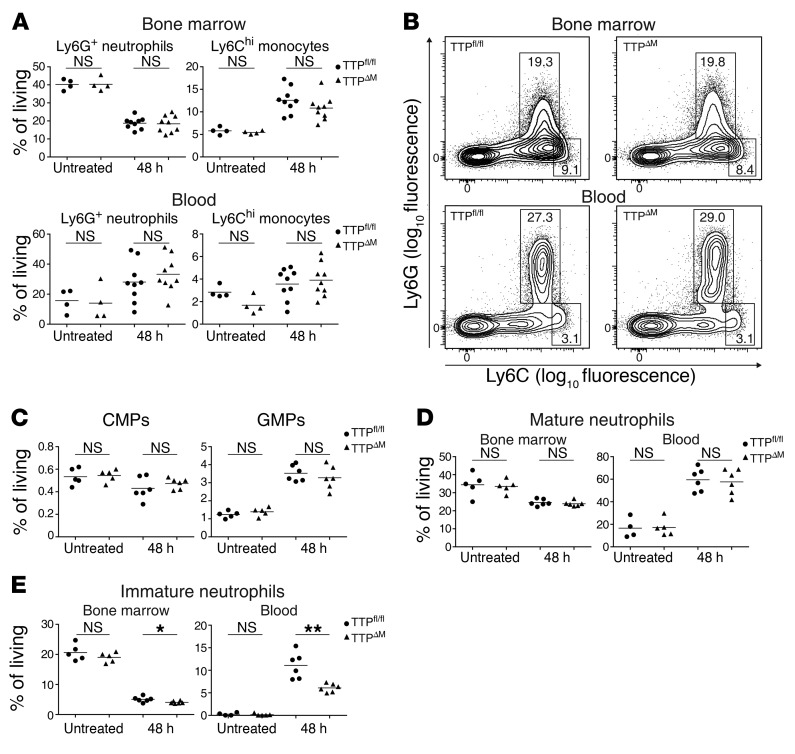
Figure 2. Bone marrow and circulating neutrophils and monocytes are not regulated by TTP during bacterial infection.
(A and B) Neutrophil and inflammatory monocyte populations in the bone marrow and blood of TTPfl/fl and TTPΔM animals. Bone marrow and blood of untreated or infected animals were analyzed by flow cytometry 48 hours p.i. Cells were subgated for CD11b+Siglec-F–FcεRI– cell populations, and neutrophils and inflammatory monocytes were detected as Ly6G+Ly6C+ or Ly6G–Ly6Chi cells, respectively. Dot plots of neutrophil and inflammatory monocytes in bone marrow (A, upper panel) and blood (A, lower panel) of untreated (n = 4/genotype) and infected animals (n = 9/genotype). Representative flow plots of bone marrow (B, upper panel) and blood (B, lower panel) 48 hours p.i. Numbers indicate the percentages in the outlined area. (C) CMPs (Lin–Sca-1–c-KithiCD16/32loCD34+) and GMPs (Lin–Sca-1–c-KithiCD16/32hiCD34+) in the bone marrow of untreated and infected (48 hours p.i.) TTPfl/fl and TTPΔM mice were analyzed by flow cytometry. (D) Mature neutrophils (c-Kit–Ly6G+) and (E) immature neutrophils (c-Kit+Ly6G+) in the bone marrow and blood of untreated and infected (48 hours p.i.) TTPfl/fl and TTPΔM mice were analyzed by flow cytometry. n = 5 untreated TTPfl/fl; n = 5 untreated TTPΔM mice; n = 6 infected TTPfl/fl mice; and n = 6 infected TTPΔM mice were used in C, D, and E. Error bars represent the mean. *P < 0.05 and **P < 0.01, by unpaired Student’s t test.

