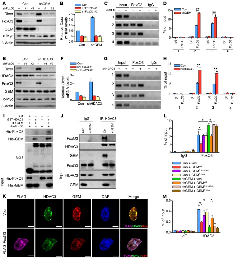
Figure 3. Geminin suppresses Dicer expression via coupling HDAC3 to FoxO3.
(A and B) Whole cell lysates prepared from LM2 cells infected with lentivirus encoding the indicated shRNAs were subjected to Western blotting analysis with indicated antibodies (A) and qRT-PCR analysis of Dicer mRNA levels (B). (C and D) Anti-FoxO3 or control IgG was used for ChIP assay to analyze the occupancy of FoxO3 on the Dicer promoter in LM2 cells with control versus geminin depletion (C). Quantification results are shown (D). n = 3 independent experiments. **P < 0.01, 2-tailed Student’s t test. (E and F) LM2 cells infected with lentivirus encoding the indicated shRNAs were subjected to Western blotting analysis with the indicated antibodies (E). qRT-PCR analysis of Dicer mRNA levels are shown (F). (G and H) ChIP assay to analyze the occupancy of FoxO3 on Dicer promoter in control versus HDAC3-depleted LM2 cells (G). Quantification results are shown (H). n = 3 independent experiments. **P < 0.01, 2-tailed Student’s t test. (I) Purified geminin and/or FoxO3 recombinant proteins were incubated with GST or GST-HDAC3. Proteins retained on sepharose were then blotted with the indicated antibodies. (J) Lysates from LM2 cells expressing geminin shRNA (shGEM) were subjected to immunoprecipitation with HDAC3 antibody followed by immunoblotting with anti-FoxO3, anti-HDAC3, and anti-geminin. (K) LM2 cells were infected with lentivirus-encoding vector or FLAG-FoxO3 for 72 hours. Cells were then fixed and stained with the indicated antibodies. Colocalization of the indicated proteins was visualized by confocal microscope. Scale bars: 10 μm. (L and M) ChIP to analyze the occupancy of FoxO3 (L) or HDAC3 (M) on Dicer promoter in geminin-depleted LM2 cells reconstituted with the indicated geminin constructs. n = 3 independent experiments. *P < 0.05, 2-way ANOVA with Bonferroni’s post hoc test.

