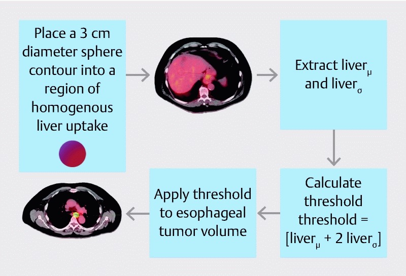
Fig. 1.

Method used to delineate an MTV threshold for each tumor. To account for background uptake, a 3-cm spherical region-of-interest is placed in the center of the liver on the PET/CT image. This method was previously described by Venkat et al. 33 .
