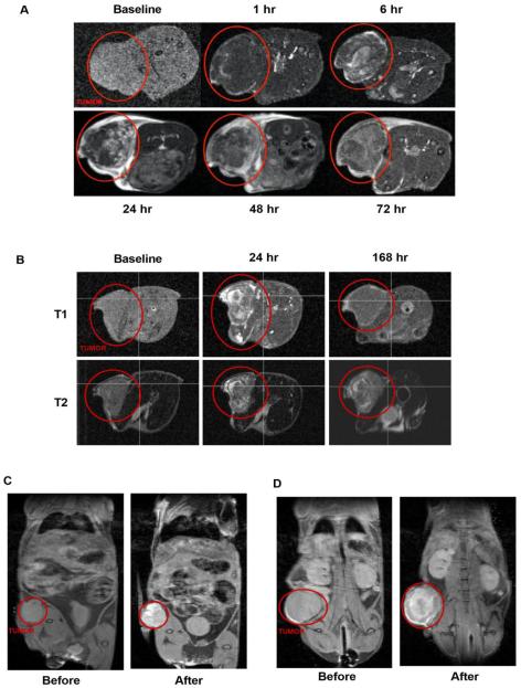
Figure 4.
MR imaging of iron-stabilized micelles in mouse subcutaneous xenograft models. (A) Time-course T1-weighted MRI of HCT116 mouse xenograft following IV administration of SN-38-loaded, iron-stabilized micelle formulation (IT-141). Tumor is identified by red circle in both panel (A) and (B). Signal peaks around 24-48 hours and is mainly cleared by 168 hours (B). Time-course T1-weighted MRI compared to T2-weighted MRI in HCT116 xenograft model. Pre-dose and 48 hours MRI image of HCT116 (C) and NCI-H460 (D) mouse xenograft following IV administration of epothilone D-loaded, iron-stabilized micelle formulation (IT-147).

