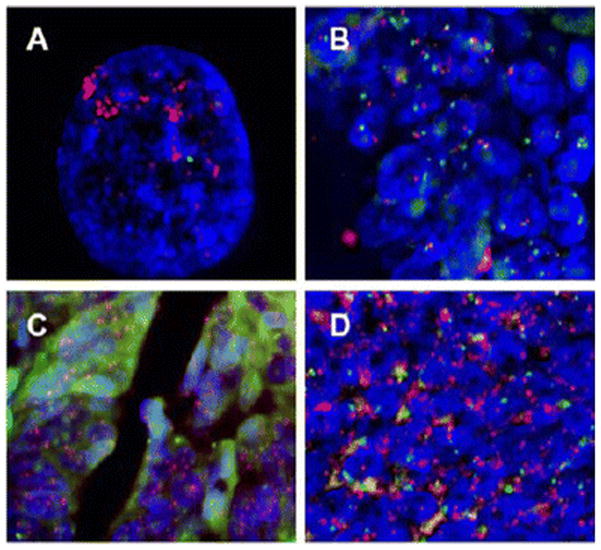
Figure 1.

Representative FISH analysis performed on formalin-fixed, paraffin-embedded human colon cancer samples. Dual-color FISH probes contain EGFR (red signals) and centromere of chromosome 7 (green signals). DAPI (blue) was used as counterstain. A shows EGFR gene amplification in human epidermoid carcinoma cell line A-431 with well-documented EGFR gene amplification. B shows colon cancer without EGFR amplification. C and D show colon cancer with EGFR amplification.
