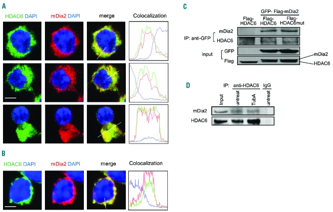
Figure 4.
mDia2 interacts with HDAC6 during erythropoiesis. Confocal microscopy analysis of HDAC6 and mDia2 co-localization in cultured Ter119-negative mouse fetal progenitors treated with (A) DMSO or (B) TubA. The florescent intensity was measured using Volocity imaging software. Scale bar is 5 μm. (C) 293T cells were transfected with GFP-Flag-tagged mDia2 and Flag-tagged HDAC6 or HDAC6 mutant as indicated. The cell lysate was immunoprecipitated with GFP antibody. Associated proteins were detected by western blotting with antibodies as indicated. (D) Endogenous HDAC6 in MEL cells treated with TubA and immunoprecipitated with HDAC6 antibody. Associated mDia2 was detected by western blotting using anti-mDia2 antibody.

