
Figure 1.
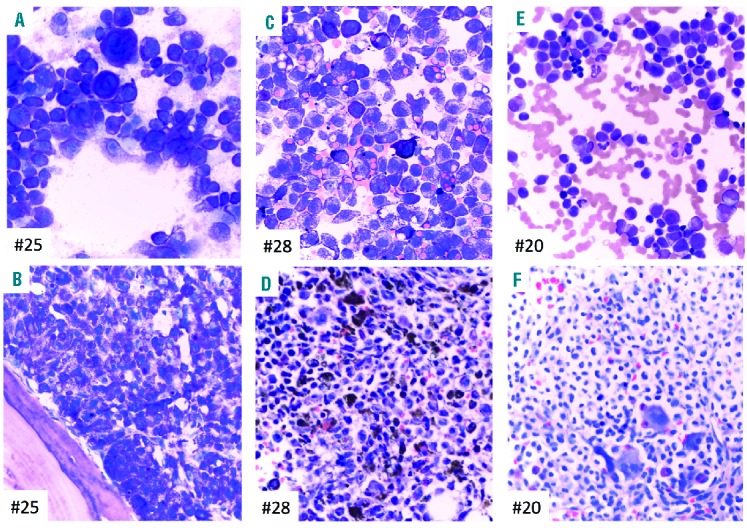
Bone marrow morphology and phenotype of 3 patients with mast cell leukemia (MCL). Patients #25: MCL, #28: MCL, #20: MCL with an associated hematologic neoplasm (AHN). (A, C and E) Bone marrow (BM) smears show an abundance of pleomorphic mast cells (MCs) exhibiting metachromatic granules. Quantitative criteria for diagnosis of MCL are completely fulfilled since MC make up more than 90% of all nucleated cells. Note the bizarre giant metachromatic MCs in (A), the prominent hemophagocytic activity of MC in (C), and the marked cytological atypia of MCs with pronounced hypogranulation in (E). Case (E) also exhibits immature atypical eosinophils enabling the diagnosis of an AHN, probably myelodysplastic/myeloproliferative neoplasm with eosinophilia. (B, D and F) BM sections show extreme hypercellularity and packed MC infiltrates. Fat cells and normal blood cell precursors are subtotally depleted. The cytomorphological aspects are fully reflected by the histomorphological findings. Note the extreme siderosis in (D) and the clear-cell aspect of atypical MC in (F) with the possibility of a misdiagnosis (hairy cell leukemia, histiocytosis or even metastatic infiltrates of a renal cell carcinoma) unless appropriate immunohistochemistry is performed. (A–F) Wright-Giemsa staining.