Figure 4.

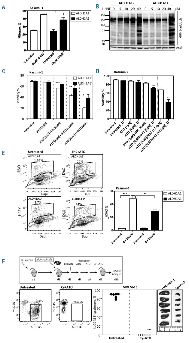
Overexpression of ALDH1A1+ partially rescues ALDH1A1− acute myeloid leukemia (AML) cell sensitivity to toxic substrates of ALDH. (A) ROS (Mitosox) detection in ALDH1A1+ and ALDH1A1- Kasumi-1 cells after six hours of treatment with 40 μM 4-HNE (n=3;*P<0.05). (B) Western blot analysis of 4-HNE protein adducts generated by treating ALDH1A1+ and ALDH1A1− Kasumi-1 cells with different concentrations of 4-HNE for 1 hour. (C) Viability of Kasumi-1 cells engineered to express ALDH1A1 through lentiviral gene transfer and treated with various combinations and doses of 4HC and ATO (n=3 replicates; ***P<0.0005, **P<0.005, *P<0.05). (D) Viability of Kasumi-3 cells treated with various combinations and doses of 4HC and ATO with (black) or without (white) the ALDH inhibitor DEAB (D) (n=3; **P<0.005, ***P<0.0005). (E) Flow cytometric assessment and summary graph of DNA damage (γH2AX) in ALDH1A1+ and ALDH1A1− Kasumi-1 cells treated with 4HC+ATO (n=3; **P<0.005, ***P<0.0005). (A–C) Bars represent mean±SD. Technical triplicates were performed. (F) In vivo treatment with Cy and ATO of NSGS mice with established ALDH1A1− MOLM-13 leukemia (n=6; ***P<0.0005).
