
Figure 7.
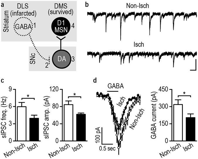
Stroke-mediated DLS infarction reduces GABAergic inputs to DMS-projecting SNc neurons. (a) Schematic of proposed changes in striatal and midbrain neurons after DLS infarction. The death of GABAergic DLS neurons (1) may disinhibit dopamine neurons (2) that project to the DMS, leading to increased activity of these neurons (3, 4). (b) Representative sIPSC traces in DMS-projecting SNc neurons on the Non-Isch and Isch sides. Scale bars: 50 ms, 100 pA. (c) Stroke-induced DLS infarction reduced the frequency and amplitude of sIPSCs in DMS-projecting SNc neurons. Left, bar graphs summarizing the average sIPSC frequencies. *p < 0.05. Right, bar graphs displaying the average amplitudes. *p < 0.05, t test. n = 12 neurons from 4 rats (Non-Isch) and 12 neurons from 5 rats (Isch) for both left and right graphs. (d) Stroke-induced DLS infarction resulted in lower GABA-induced currents in DMS-projecting SNc neurons. Left, changes in the holding current in DMS-projecting SNc neurons on the contralesional non-ischemic (Non-Isch) and ipsilateral ischemic (Isch) sides. Right, bar graphs summarizing the averaged GABA currents in SNc neurons on both sides. *p < 0.05, t test. n = 9 neurons from 5 rats per group.