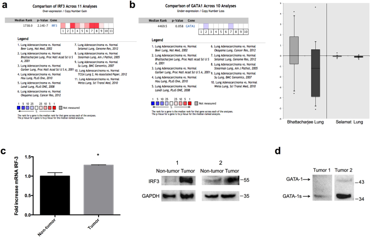
Figure 3.
Upregulation of IRF-3 protein expression and down-regulated expression of GATA-1 protein in human lung adenocarcinoma tissue. (a) Eleven analyses were evaluated in comparing the RNA expression of IRF-3 between lung adenocarcinoma and normal tissue. Values above the average were considered IRF-3 over-expression (red). (b) Ten analyses of GATA1 gene demonstrated the different expression in lung adenocarcinoma compared to normal tissue. Lower expression of GATA1 was shown in two studies (blue). These two studies, Bhattacharjee Lung and Selamat Lung datasets studies, showed GATA-1 expression in normal tissues (light grey) and lung adenocarcinoma (dark grey). Log2 Median-Centered ratio was used to average all normal samples in data analysis. (c) The endogenous IRF-3 mRNA and protein expression was examined in lung adenocarcinoma and adjacent normal tissue. P values were determined by Student’s-t test (*p < 0.05). (d) The GATA-1 protein expression was detected in lung adenocarcinoma tissue by Western blot. GAPDH was used as a internal control. The full-length blots were presented in Supplementary Figure S2.

