Figure 3.

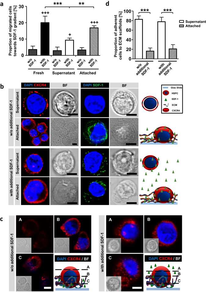
SDF-1/CXCR4 axis is active on ECM cultured HSPCs. (a) Proportion of migrated fresh-, SN- and AT-cells in a trans-well migration assay towards SDF-1 containing medium (with SDF-1) or towards SDF-1 free medium (spontaneous migration, w/o SDF-1). n = 4, two-tailed t-test; * = significance in comparison to SN-cells and + = significance in comparison to corresponding w/o SDF-1. (b) Confocal microscopy images of CXCR4 (red) or SDF-1 (green) staining and nuclear DAPI (blue) staining. Cells were treated with medium either without (w/o) or with additional SDF-1. Schemas show technique and findings. Bars = 5 µm. (c) Confocal microscopy z-stack images of α-CXCR4 (red) and nuclei DAPI (blue) staining. A = top, B = middle and C = bottom of cells. Cells were treated with medium either without or with additional SDF-1. Schemas show technique and findings. Bars = 5 µm. (d) Proportion of cell adhesion to ECMs 24 hours after seeding either without or with additional SDF-1 containing medium. n = 6 for w/o additional SDF-1, n = 3 for with additional SDF-1, two-tailed t-test. Error bars, SD.; *p < 0.05, **p < 0.01, ***p < 0.001.
