Figure 4.

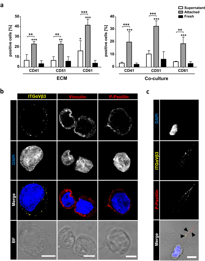
Integrins recognizing RGD-motives promotes active focal contact formation. (a) FACS analysis of freshly isolated cells or 5 days ECM or MSC co-cultured cells. Percent positive cells for ITGαIIb (CD41), ITGαV and ITGβ3 (CD61) are shown. n = 4, two-way ANOVA with Bonferroni post-hoc test; + = significance in comparison to fresh cells. Error bars, s.e.m.; *p < 0.05, **p < 0.01, ***p < 0.001. (b) Confocal microscopy images of α-ITGαVβ3 (left panel, yellow), α-vinculin (mid penal, red), α-p-paxillin (right panel, red), nuclei DAPI (blue) staining, merge and corresponding brightfield images of 5 days cultured AT-cells. Bars = 5 µm. (c) Confocal microscopy images of α-ITGαVβ3 (yellow), α-p-paxillin (right panel, red), nuclei DAPI (blue) staining and merge including corresponding brightfield image of 5 days cultured AT-cell. Arrowheads represent co-localization of ITGαVβ3 and p-paxillin in a cellular protrusion. Bar = 5 µm.
