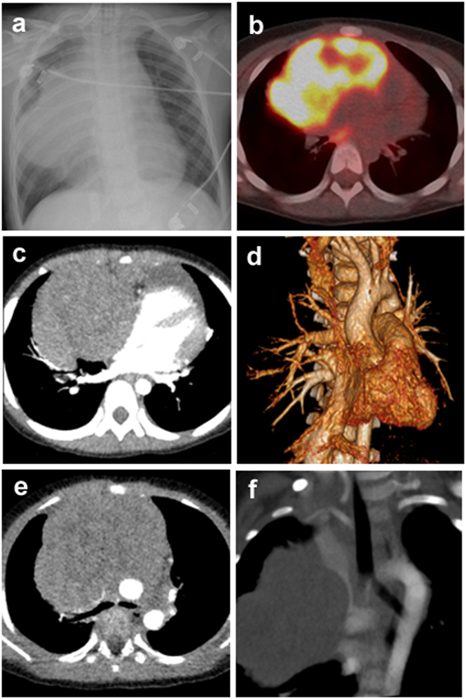
Figure 1.

Radiological characteristics of T-LBL/T-ALL involving the mediastinum, case 1. (a) Chest X-ray image shows a large mass in the mediastinum. (b) PET-CT image shows increased FDG activity in the mediastinum. (c) Axial contrast-enhanced CT scan shows the compression of pericardiaum. (d) Three-dimensional volume rendered image of contrast-enhanced CT exhibits the compression of pericardiaum. (e) Axial contrast-enhanced CT scan shows vascular compression and tracheal encasement of the mass. (f) Maximal intensity projection of contrast-enhanced CT reveals the encasement of superior vena cava.
