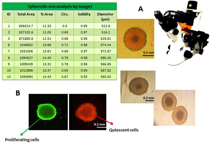
Figure 2.

MDA-MB-231 cell spheroids. (A) MDA-MB-231 cell spheroid’s size and morphology inspected and imaged by bright field invert microscope. (B) AO and EB staining of a uniform spheroid, dark and orange highlighted regions (stained with EB) close to the center of the spheroid are mainly composed of quiescent/dead cells whereas, outermost layer which is highlighted in bright green (stained with AO) consists of proliferative, actively dividing cells.
