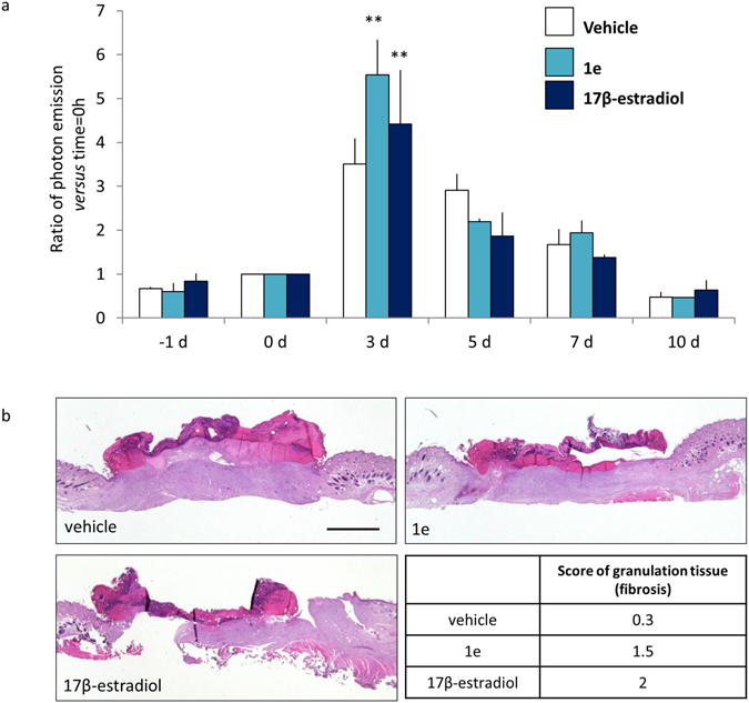
Figure 10.

1e compound and 17β- estradiol accelerate wound healing. (a) In vivo BLI: round wound area photon emission quantification at indicated time point in MITO-Luc mice. Each bar represents the mean ratio versus time = 0 h +/− SEM of photon emission measured in 2 animals/group. (**p < 0.01 vs 0 h, ANOVA followed by Bonferroni’s test for multiple comparisons). (b) Hematoxylin-eosin representative staining of wound section at 10 days after the indicated treatments. The histoscore value for granulation tissue (fibrosis) is shown as mean, each histoscore consists of 4 random fields/groups, the criteria for the assignation of histoscore value are: 1 = wound bed partially covered with granulation tissue; 2 = thin granulation over the whole wound bed; 3 = thick granulation over the whole wound bed. Bar = 2 mm.
