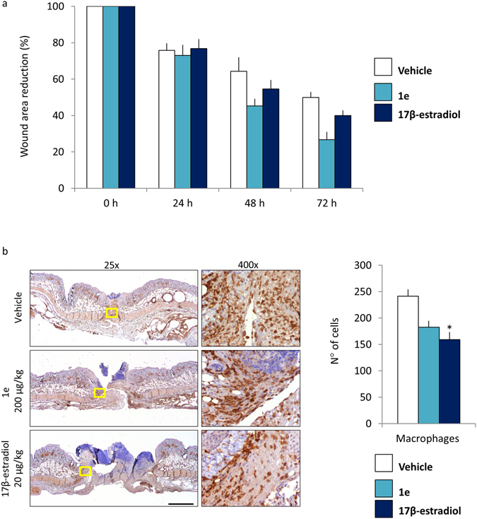
Figure 9.

Reduction of the wound areas after treatment with 1e and 17β-estradiol. (a) Wound area measurement: each bar represents the mean ratio versus time = 0 h +/− SEM expressed as percentage of wound area measured in 4 animals/group. (b) Representative staining of wound explants at 72 hours after treatments. Staining was done with Iba1 antibody. Pictures were taken at 25x and 400x. Bar = 2 mm. The number of macrophages cells around wound site: each bar represents the mean +/−SEM, macrophages (Iba1 positive cells) were counted by analyzing n = 6 fields (vehicle), n = 11 fields (1e) and n = 10 fields (17β-estradiol) at 400x enlargement. (*p < 0.1 vs vehicle, ANOVA followed by Bonferroni’s test for multiple comparisons).
