Figure 6.

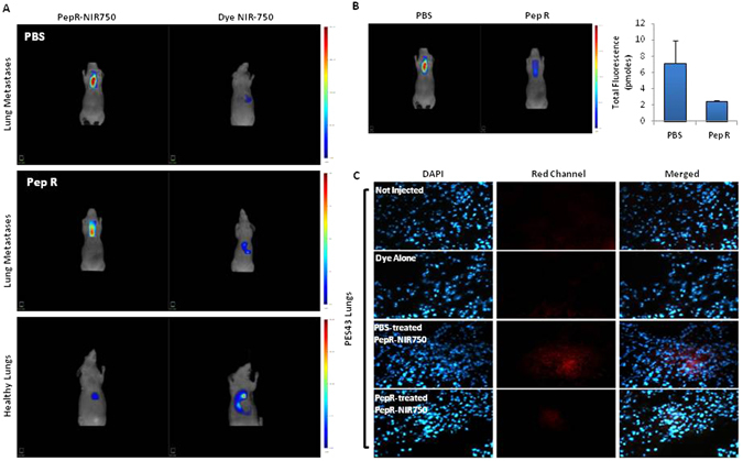
PepR-NIR750 detects lung metastases reduction induced by Peptide R treatment. CD-1 nu/nu athymic mice bearing PES43-derived lung metastases were treated with PBS (control group) or Peptide R (2 mg/Kg) 5 days/week for 2 weeks. 3 weeks later mice were i.v. injected with PepR-NIR750 or VivoTag-S 750 alone and total body imaged after 24 h with FMT4000. (A) PepR-NIR750 signal is detectable in mice bearing lung metastases (upper and middle left panels) while no signal was revealed with VivoTag-S 750 alone (upper and middle right panels). Healthy lungs are not imaged by PepR-NIR750 (lower panel). (B) In vivo imaging of mice bearing lung metastases treated and not treated with Peptide R using Pep-R-NIR750. Peptide R causes a strong reduction of PepR-NIR750 signal (65%) respect to control group as observed 24 hours after its injection. (C) After 24 h the mice were euthanized, the lungs snap frozen and then were 5 µm sliced at cryostat microtome. Slides of PepR-NIR750 or VivoTag-S 750 were imaged at fluorescence microscope Axioscope A.1.
