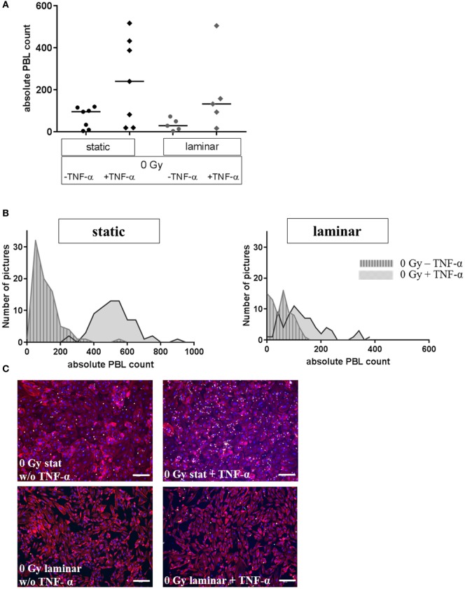
Figure 2.
Adhesion assay of PBL to stimulated human microvascular endothelial cell (HMVEC) under static or laminar flow culture conditions. Adhesion of PBL to HMVEC is shown under static or laminar culture conditions, treated with or w/o TNF-α. Absolute counts of adherent PBL to HMVEC are shown in (A); N static = 7, N laminar = 5; duplicates or triplicates were used for each experiment; line = median. Distributions of adherent PBL counts are shown in (B) for one representative experiment each; left: static culture conditions, right: laminar culture conditions; gray/striped: untreated, gray: with TNF-α. Representative microphotographs are given in (C). Upper panel: static culture conditions, without (left) or with TNF-α (right); lower panel: laminar culture conditions, without (left) or with TNF-α (right). Blue = DAPI/nuclei, red = TRITC-Phalloidin/cytoskeleton, green = CFDA/PBL; 100×, bars = 100 µm.

