Figure 4.
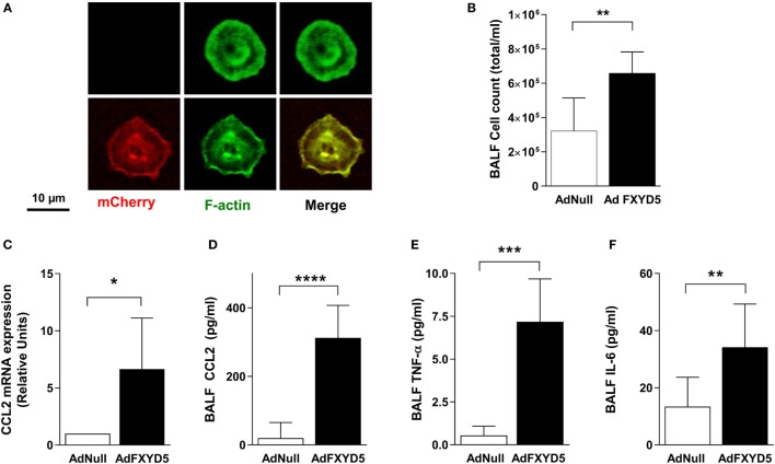
Increased levels of FXYD5 are sufficient to induce lung inflammation. Mice were instilled with adenoviruses encoding m-Cherry-HA-FXYD5 (AdFXYD5) or an empty control (AdNull) and 72 h later ATII cells, BALF or lung peripheral tissue were isolated. (A) Confocal microscopy analyses of FACS-sorted mice ATII cells. Red m-cherry fluorescence reflects the expression of FXYD5. Green fluorescence shows actin filaments used to visualize cells. (B) Total cell count in BALF. (C) CCL2 mRNA was determined by RT-qPCR in lung peripheral tissue. (D–F) CCL2, TNF-α, and IL-6 were determined by ELISA in BALF. Values of Ad-Null-treated controls were normalized to 1. Bars represent means ± SD n ≤ 5. Statistical significance was analyzed by unpaired Student’s t-test. *p ≤ 0.05; **p ≤ 0.01; ***p ≤ 0.001.

