Fig. 1.

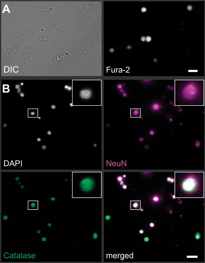
Primary dissociated cell culture and fura-2 imaging of nucleus tractus solitarii (nTS) neurons. A: example of nTS cells visualized using differential interference contrast (DIC; left) also show strong fura-2 fluorescence when excited with 380 nm (right). Scale bar = 20 µm. B: immunocytochemical verification on paraformaldehyde-fixed nTS cells that our cultures primarily consisted of neurons (NeuN, magenta) that colabeled with nuclei (DAPI, gray) and the antioxidant catalase (green). Scale bar = 20 µm. Inset: zoomed image of one nTS neuron.
