
Fig. 4.
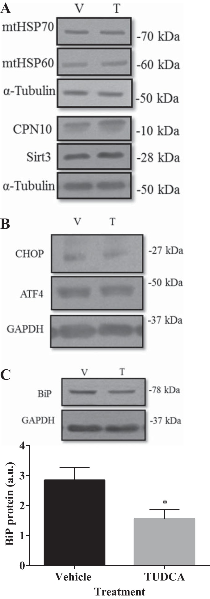
TUDCA treatment decreased BiP protein but had no impact upon the UPRMT after 4 days of skeletal muscle differentiation. Cells were pretreated for 24 h in 500 µg/ml of TUDCA (T) or water [vehicle (V)] before differentiation for 4 days. Representative Western blots of UPRMT proteins mtHSP70, mtHSP60, CPN10, and Sirt3 (A) were all similar in protein expression between vehicle- or TUDCA-treated cells. No effect of TUDCA was observed for UPRER markers CHOP and ATF4 (B). C: representative blot and graphical quantification of BiP protein. α-Tubulin served as the loading control. Data are represented as means ± SE and are measured in arbitrary units (AU). *P < 0.05 vs. vehicle; n = 3 or 4 experiments.