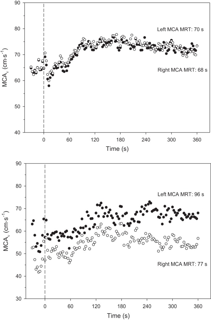
Fig. 2.

Middle cerebral artery velocity (MCAV) at rest and response following the onset of moderate-intensity exercise (dashed vertical line, time 0). Solid symbols are left MCA, and hollow symbols are right MCA. Top: illustrates subject 10 in whom there was excellent agreement between left and right. Bottom: represents poorest agreement among the young participants, subject 7. MRT, mean response time. Overall left and right MCAV correlation was 0.819 (P < 0.05) with a coefficient of variation of 7.6%.
