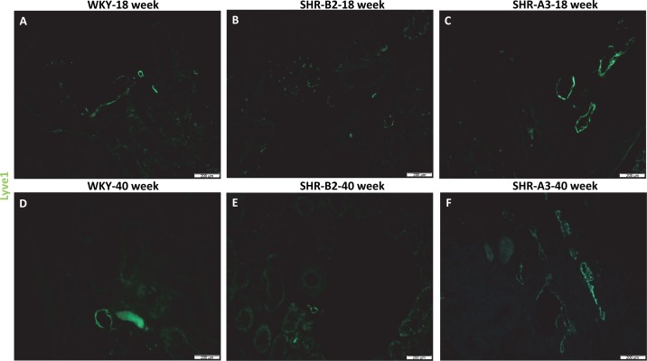
Fig. 2.
LYVE1-positive lymphatic vessels in kidneys from young and aged WKY, SHR-B2, and SHR-A3 rats. A–F: representative images show green LYVE-1 staining of lymphatic vessels in kidneys from 18-wk-old and 40-wk-old WKY, SHR-B2, and SHR-A3 rats. Similar to Fig. 1, subjectively there are reduced numbers of LYVE-1+ lymphatic vessels in SHR-B2 rats but increased size and numbers in SHR-A3 rats at 18 wk and 40 wk of age. Magnification is ×10, and the scale bar = 200 µm. The numbers of animals in each group were as follows: WKY-18 wk = 3, WKY-40 wk = 3, SHR-B2-18 wk = 3, SHR-B2-40 wk = 3, SHR-A3-18 wk = 3, SHR-A3-40 wk = 3.

