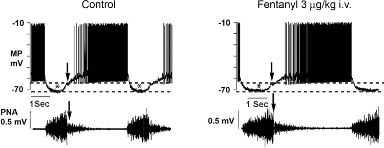
Fig. 3.
Records of a bulbospinal augmenting expiratory neuron (AugE) discharge (MP) and phrenic nerve activity (PNA) under control conditions and in response to a threshold dose of fentanyl. Dashed lines denote the amplitudes of waves (*) of inhibitory synaptic potentials that coincide with phrenic nerve inspiratory phase discharges (PNA). Arrows denote the termination of the inspiratory phase.

