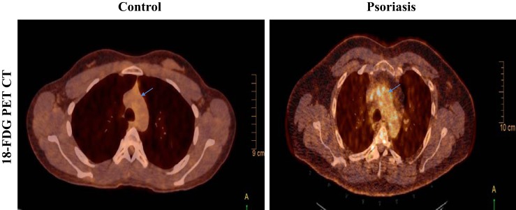
Fig. 2.
Representative images from 18fluorodeoxyglucose-positron emission tomography computed tomography (18FDG-PET/CT) scans in a healthy volunteer control (left) and a patient with psoriasis (right) exhibiting increased uptake of 18FDG at the level of aortic arch (blue arrow) as seen in the transverse section images. The images shown here are from the fused PET scans.

