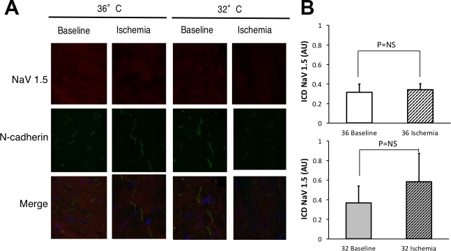
Fig. 8.
NaCh at the ID during ischemia. A: canine wedges under CT nonischemic (36° baseline, n = 3 subjects) or 15 min of ischemia (36° ischemia, n = 3 subjects) conditions and MH nonischemic (32° baseline, n = 2 subjects) or 15 min of ischemia (32° ischemia, n = 3 subjects) conditions were stained for total Nav1.5 (red) and the ID protein N-cadherin (green). Merged images show N-cadherin overlapped with total Nav1.5 images. B: summary data. There was no difference in NaCh at the ID either during ischemia (P = not significant) and no difference between temperatures (P = not significant).

