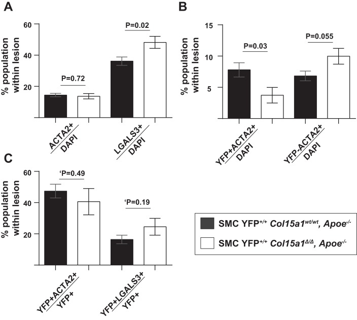
Fig. 6.
SMC-derived cell populations were reduced with SMC Col15a1 knockout. A: no changes were observed in the percentage of ACTA2+/DAPI+ cells but a significant increase was found in the percentage of LGALS3+/DAPI+ cells in SMC YFP+/+ Col15a1Δ/Δ, Apoe−/− (n = 13) compared with SMC YFP+/+ Col15a1wt/wt, Apoe−/− (n = 11) lesions. B: a significant decrease in SMC-derived ACTA2+ (YFP+ACTA2+/DAPI+) cells and a near significant increase in non-SMC-derived ACTA2+ (YFP−ACTA2+/DAPI+) cell populations were also found in SMC Col15a1 knockout compared with wild type. C: no significant differences were seen in the proportion of SMCs able to express ACTA2 (YFP+ACTA2+/YFP+) or LGALS3 (YFP+LGALS3+/YFP+) as a consequence of SMC Col15a1 knockout. Values represent the average of three locations across the BCA for each genotype. Values represent means ± SE. ‘P value was determined by an unpaired two-tailed t-test analysis with Welch’s correction.

