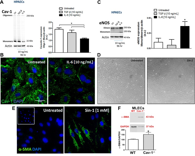
Fig. 4.
IL-6-induced Cav-1 depletion and eNOS uncoupling associated with endothelial cell reprogramming. Cav-1 expression by Western blotting (A) or immunocytochemistry (green; B) and eNOS dimer/monomer expression (C) were measured in human pulmonary artery endothelial cells (HPAECs) after treatment with TGF-β and IL-6/IL-6R for 96 h. D: phase contrast micrograph showing HPAEC morphology after SIN-1 (peroxinitrite donor) treatment for 96 h. E: α-SMA expression in murine lung endothelial cells (MLECs) from WT and Cav-1−/−. *P < 0.01 by one-way ANOVA followed by post hoc Newman-Keuls test (n = 4 cultures; passage 4–7).

