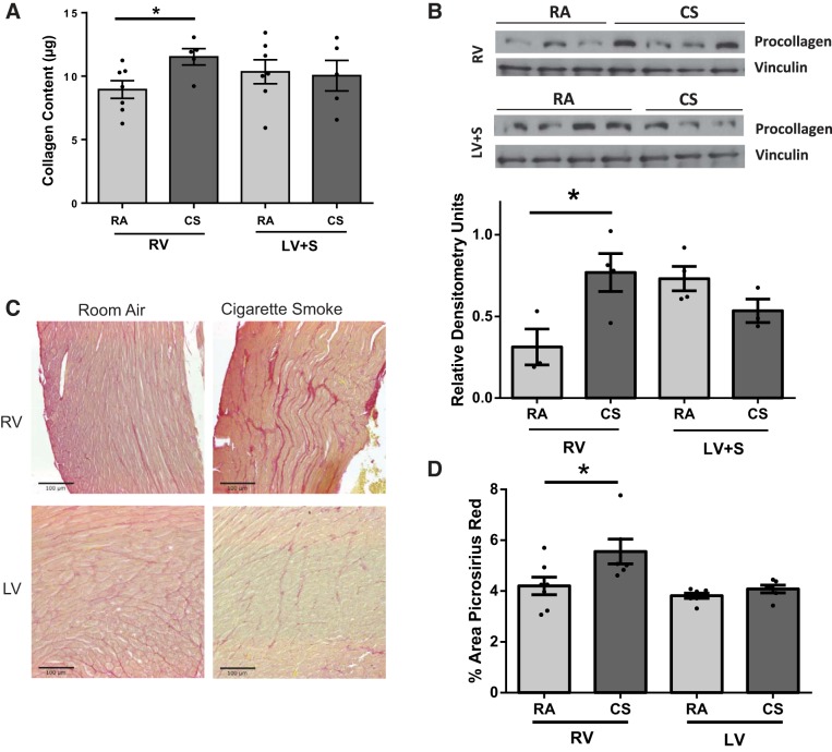
Fig. 2.
Analysis of cardiac fibrosis in RV and LV of RA- and CS-exposed mice. RV and LV + septum (S) tissue of mice exposed to CS exhibited increased collagen content (Sircol assay, per 100 μg of protein) (A) and procollagen expression (B). Sircol assay n = 5–7, procollagen expression n = 3 to 4. *P < 0.05 vs. RA, Student’s t-test. Similar increases in fibrosis were apparent with Picrosirius red-stained paraffin-embedded sections. C: representative images of RA- and CS-exposed RV and LV. D: significantly increased Picrosirius staining in CS-exposed RV but not LV, as determined by quantitation of images. *P < 0.05 vs. RA, Student’s t-test, n = 6 to 7. Scale on images = 100 μM.

