Fig. 4.
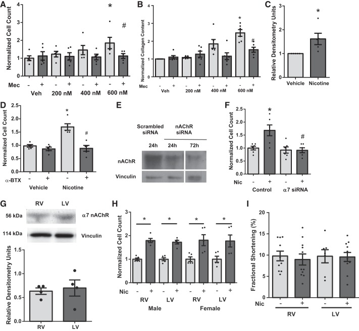
Effect of nicotine on isolated cardiac fibroblast proliferation and dependence of α7 nAChR. Nicotine increases cell counts (n = 5) (A) and collagen content (n = 5) (B) in cardiac fibroblasts through nAChR activation that was inhibited by Mec (20 μM). C: nicotine also increases procollagen expression in cardiac fibroblasts (n = 5). D: α-bungarotoxin (α-BTX; 100 nM) blocks nicotine-induced proliferation (n = 5). Knockdown of α7 nAChR with siRNA (E) blocks nicotine-induced proliferation (F) (n = 5). Quiescent cells were pretreated with either vehicle, Mec, α-BTX, or α7 nAChR siRNA, followed by vehicle or nicotine (600 nM) for 24 h. Data are normalized to vehicle treatment and presented as means ± SE. *P < 0.05 compared with Veh; #P < 0.05 compared with Mec/vehicle. G: there were no differences in expression of α7 nAChR in rat LV and RV tissues (n = 4). H: there were no changes in response to nicotine (Nic) in cardiac fibroblasts isolated from LV or RV or male or female rats. Nicotine significantly increased proliferation in all groups compared with respective untreated groups (*P < 0.05) (n = 3–4 animals/group performed in triplicate, 3-way ANOVA, Student-Newman-Keuls post hoc). I: there were no significant changes (t-test) in RV or LV cardiomyocyte contractile function with acute nicotine treatment.

