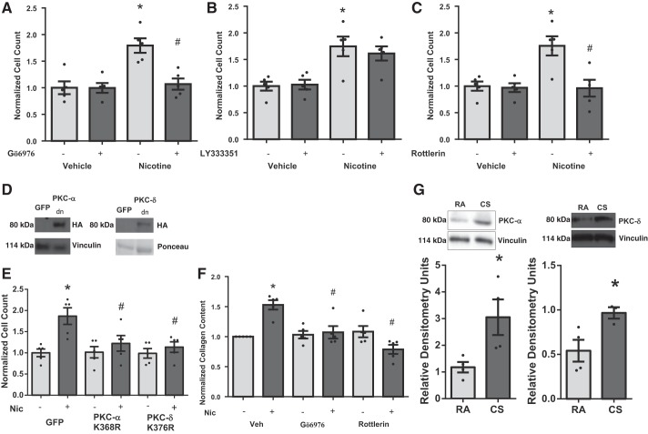
Fig. 5.
Role of PKC signaling in nicotine-induced cardiac fibroblast proliferation. Small-molecule inhibitors of PKC-α (100 nM) (A) or -δ (3 μM) (C), but not PKC-β (50 nM) (B), block nicotine-induced cardiac fibroblast proliferation (n = 5). Expression of dominant-negative (Κ368Ρ) PKC-α (D) or dominant-negative (K376R) PKC-δ (E) also blocks nicotine-induced cardiac fibroblast proliferation (n = 5). GFP, green fluorescent protein; HA, hemagglutinin. Chemical inhibition of PKC-α and -δ blocks nicotine-induced increased collagen content (F). Cells were quiesced in serum-free medium for 24 h before nicotine stimulation. *P < 0.05 vs. Veh/Veh, and #P < 0.05 vs. Veh/Nic using 2-way ANOVA, Student-Newman-Keuls post hoc test. CS-exposed mice have increased expression of PKC-α and -δ in RV tissue. G, top: representative blot is shown. Bottom: quantitation of data in G (n = 4). *P < 0.05 vs. RA.

