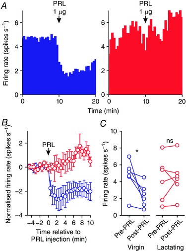
Figure 5. Prolactin effects on the activity of oxytocin neurones in virgin and lactating rats after bromocriptine treatment.

A, example ratemeter recordings of oxytocin neurone firing rate (in 30 s bins) from urethane‐anaesthetised bromocriptine‐treated virgin (left) and lactating (right) female Sprague–Dawley rats, showing that i.c.v. administration of 1 μg prolactin (PRL) reduces firing rate in the neurone from the virgin rat but not in the neurone from the lactating rat. B, mean (±SEM) oxytocin neurone firing rate (in 30 s bins) before and after i.c.v. administration of 1 μg prolactin in virgin rats (blue symbols) and lactating rats (red symbols). C, mean oxytocin neurone firing rate averaged over 5 min before and after i.c.v. administration of 1 μg prolactin in virgin rats (left panel; n = 6) and lactating rats (right panel; n = 6); * P < 0.05 (t 5 = 2.89) and ns P > 0.05 (t 5 = −0.37) vs. pre‐prolactin, paired t test. [Color figure can be viewed at wileyonlinelibrary.com]
