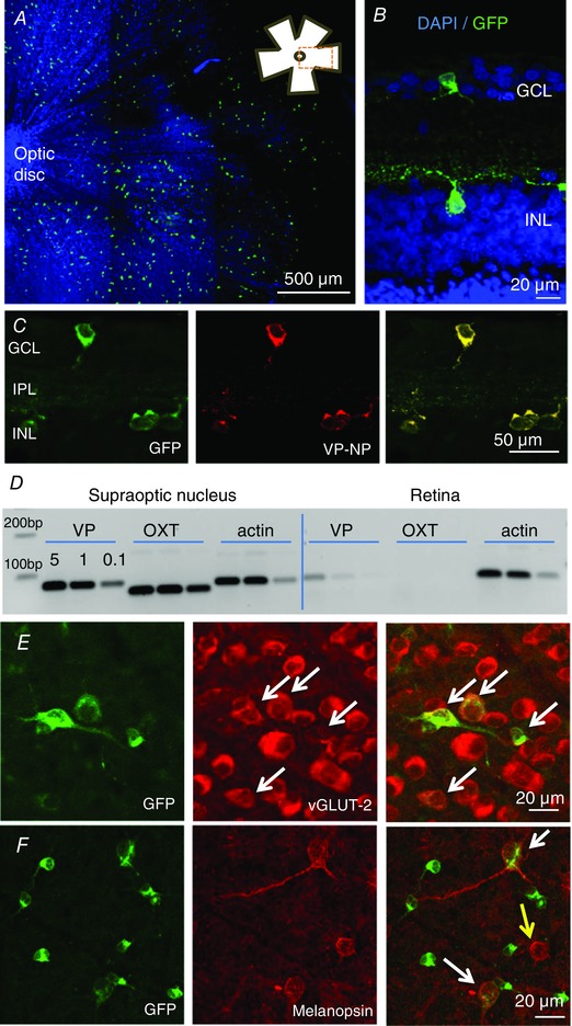
Figure 1. Vasopressin neurons in the retina.

A, flat mount of retina, focused on the ganglion cell layer (GCL), shows dispersed eGFP‐expressing cells (green cells); the blue staining is a nuclear marker DAPI, to show the location of all cells in the field of view; the inset shows the location of the image within the flat mount. B, eGFP‐cells occur in both the ganglion cell layer (GCL) and the inner nuclear layer (INL), as shown in a cross section of the flat mount. C, eGFP‐cells express vasopressin–neurophysin (VP‐NP); the successive images show fluorescence for eGFP, immunoreactive VP‐NP and overlaid images. D, PCR confirmation of expression of vasopressin mRNA (VP, 77 bp) and actin mRNA in the supraoptic nucleus (positive control) and the retina of wild‐type rats; the supraoptic nucleus contains magnocellular neurons that project to the posterior pituitary gland. Note there is no detectable oxytocin mRNA (OXT, 62 bp) in the retina; oxytocin is a closely related peptide that is also expressed in the supraoptic nucleus. E, eGFP‐cells (shown in a flat mount) co‐express the vesicle glutamate transporter vGLUT‐2 (white arrows) indicating that they use glutamate as a conventional neurotransmitter. F, some eGFP‐cells co‐express the photopigment melanopsin (white arrows; yellow arrow shows a cell immunopositive for melanopsin only).
