
Figure 7. Expression of RXFP3 mRNA by OT and AVP PVN neurons.
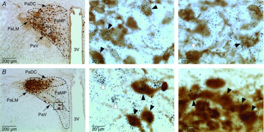
A, left, low‐power image of the paraventricular hypothalamic nucleus (PVN) of a rat, in which RXFP3 mRNA was detected by in situ hybridization histochemistry and oxytocin (OT) was detected by immunohistochemistry; middle, high‐magnification image of neurons in the ventral part of the PVN (PaV) outlined by a rectangle in the left panel; and right, high‐magnification image of neurons in the lateral magnocellular part of the PVN (PaLM) outlined by a rectangle in the left panel. Some double‐labelled neurons positive for OT‐immunoreactivity (brown staining) and RXFP3 mRNA (black silver grains) are indicated by black arrowheads. A white arrowhead in the right panel depicts an OT neuron negative for RXFP3 mRNA. B, left, low‐power image of the PVN of a rat, in which the expression of RXFP3 mRNA was detected by in situ hybridization histochemistry and arginine vasopressin (AVP) was detected by immunohistochemistry; middle, high‐magnification image of neurons in the ventral part of the PVN (PaV) outlined by a rectangle in the left panel; and right, high‐magnification image of neurons in the lateral magnocellular part of the PVN (PaLM) outlined by a rectangle in the left panel. Some double‐labelled neurons positive for AVP‐immunoreactivity (brown staining) and RXFP3 mRNA (black silver grains) are indicated by black arrowheads. Neurons expressing RXFP3 mRNA but negative for AVP‐immunoreactivity are indicated by white arrowheads. Abbreviations: 3V, third ventricle; PaDC, dorsal cap of PVN; PaLM, lateral magnocellular part of PVN; PaMP, medial parvocellular part of PVN; and PaV, ventral part of PVN. [Color figure can be viewed at wileyonlinelibrary.com]