
Figure 5. Optogenetics‐based selective activation of geniculohypothalamic signalling.
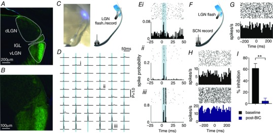
A, GFP‐immunostaining enhanced ChR2/EYFP expression in the thalamus of GAD2‐Cre; Ai32 mice, revealed robust expression in neural processes across the predominantly GABAergic IGL/vLGN region. B, central region of (A) showing EYFP signal across the dLGN and IGL/vLGN at higher magnification. C, live image of slice showing optical fibre positioning over IGL/LGN (left) and schematic of slice preparation with optical fibre and pMEA placements used (right). D and E, direct optical stimulation (465 nm LED; ∼800 mW mm−2 at fibre tip, 10 ms flash; represented by blue shaded area in traces) evoked robust and rapid excitatory responses across the IGL/vLGN and much weaker responses in the dLGN. Traces in (D) represent the probability of spike occurrence (from 200 trials) relative to stimulation onset at each site on the 59‐electrode recording array for the slice shown in (C). Data in (Ei, Eii and E iii) show perievent rasters and larger spike probability histograms for the traces indicated in (D). F, schematic of slice preparation showing optical fibre and pMEA placements used for investigation of GHT signalling. G, optical stimulation of GABAergic GHT projection neurons evoked modest inhibitory responses in a small subset of SCN recording sites (n = 12 sites from four slices; inhibition, mean ± SEM: 43.8 ± 5.5% of pre‐stimulation; latency, median ± SD: 60 ± 25 ms: representative SCN recording sites are shown in G and H). H and I, where tested (n = 6), optogenetic‐evoked inhibitory responses were abolished by bath application of the GABAAR antagonist, BIC (20 μm). H, data from a representative SCN recording site pre‐ and post‐BIC (upper and lower, respectively). I, mean ± SEM response magnitude pre‐ and post‐BIC (n = 6). Data were analysed by paired t test. ** P < 0.01.