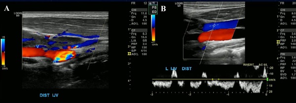
Figure 2.

Ultrasound of the neck: (A), intraluminal echogenic filling defect within the distal left jugular vein which was non-compressible, consistent with a non-occlusive thrombus. (B), repeat ultrasound after the initiation of therapy demonstrating very minimal residual thrombosis of the distal IJV.
